|
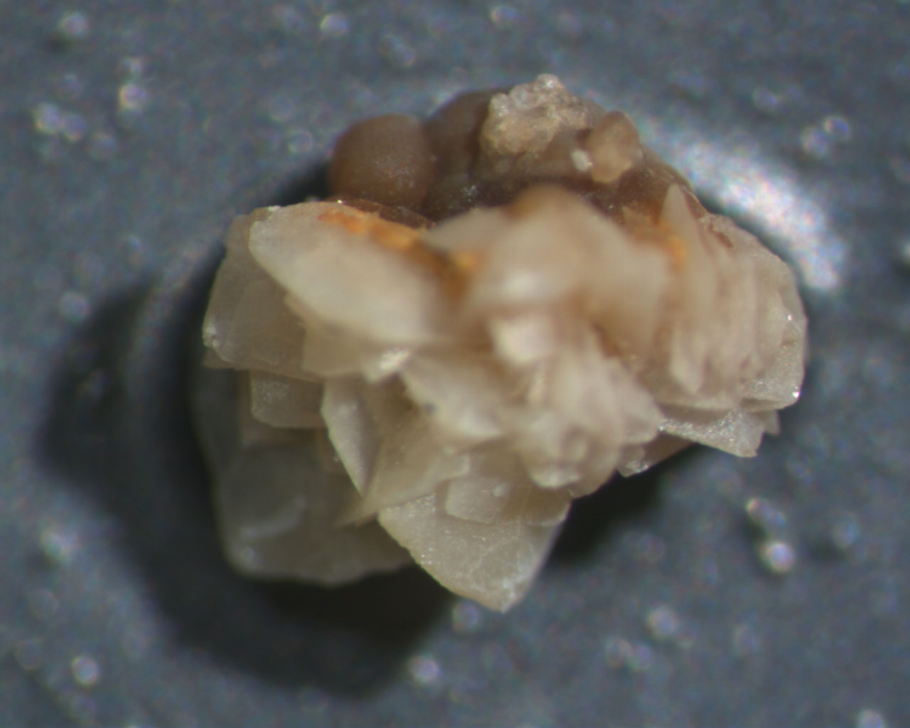
The image below is the kidney stone as seen through a dissecting microscope. Note the different colors and crystal shapes.

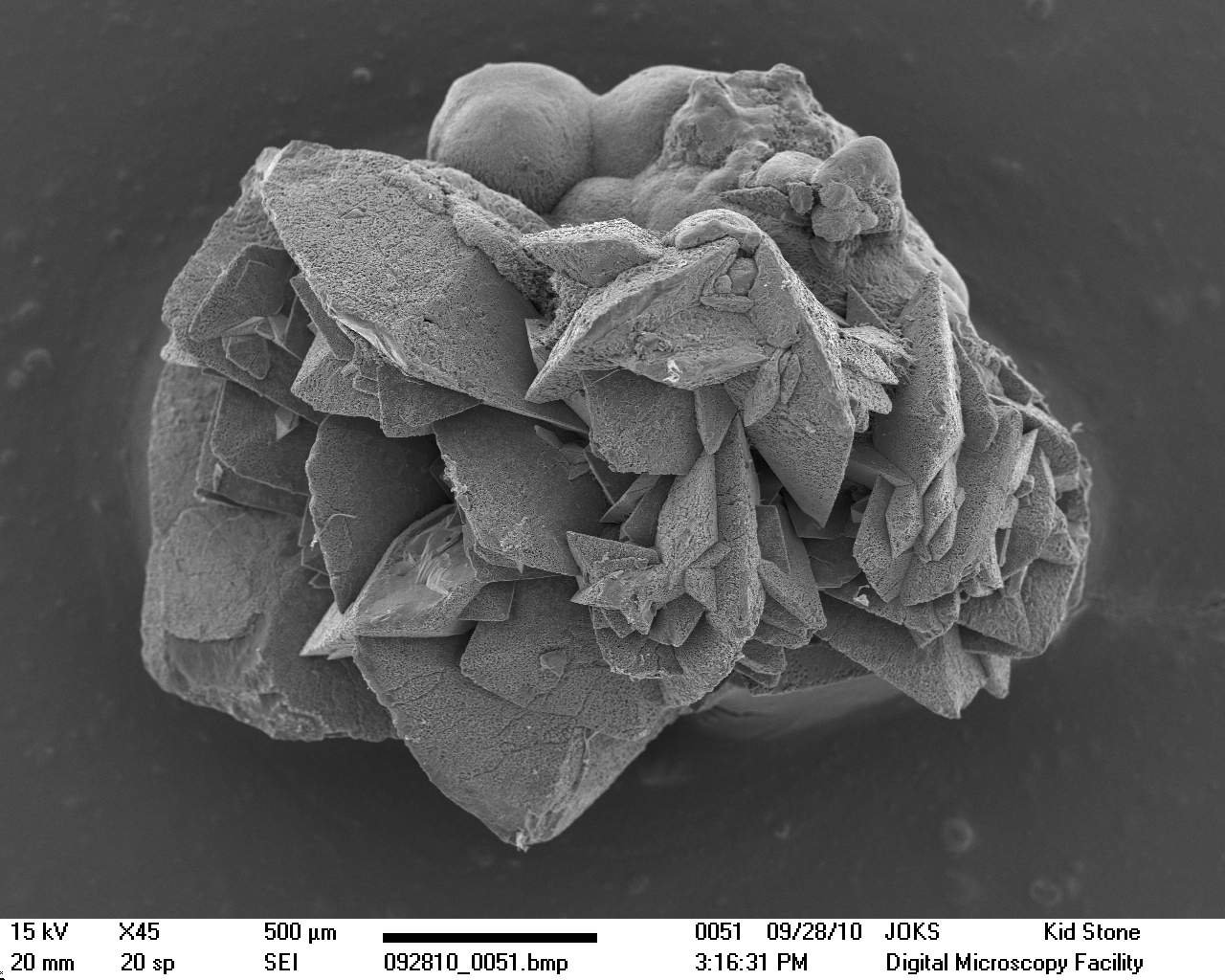
The same kidney stone coated with gold and viewed with the SEM. A collection of higher magnification views of the various crystal types is here.

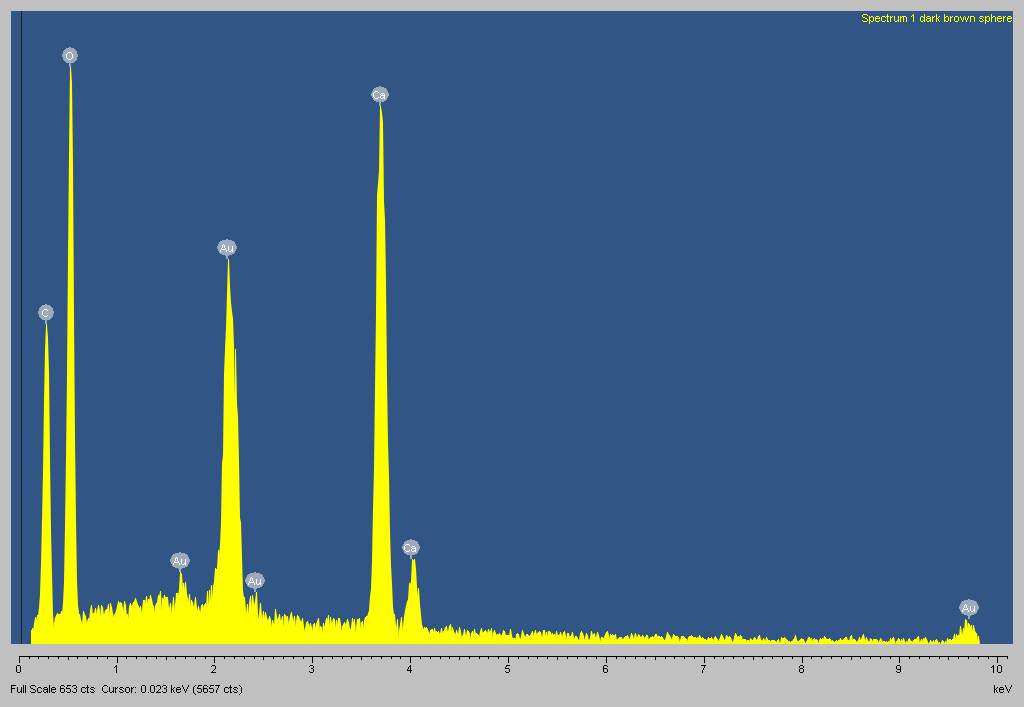
Despite the differences in color and crystal shape, EDS analysis shows that all of the material making up the stone is most likely calcium oxalate (CaC2O4) the composition of the majority (75-85%) of all kidney stones. EDS spectra from all areas of the stone were similar to that shown below. Note prominent peaks for carbon (C), oxygen (O) and calcium (Ca). The gold peaks (Au) result from the conductive gold coating

|